What Is An Echocardiogram?
An echocardiogram is a test that uses sound waves to examine, measure and produce images of the heart for diagnostic purposes. It is used by doctors to see how your heart is beating and pumping blood. He also uses it to identify abnormalities in the heart muscle and valves. It can also detect congenital heart defects in unborn babies.
 Getting An Echocardiogram |
What Can A Echocardiogram Show?
- Heart Size.
- The Pumping Strength Of Your Heart.
- Damage To The Heart Muscle.
- Heart Valve Problems.
- Heart Defects.
Why Do I Need An Echocardiogram?
You may need an echocardiogram if your doctor suspects a problem with your heart. It may be a problem with your hearts ability to pump blood or with the valves or chambers of your heart. Expect the process to take about half and hour. Expect the room to be cold and you to be a little nervous. Expect the technician to dim the lights in the room to see the monitor better. Expect to hear the sounds of the machine recording the blood flowing in your heart. Expect to be asked to hold your breath occasionally or slow your breathing to get a better picture.
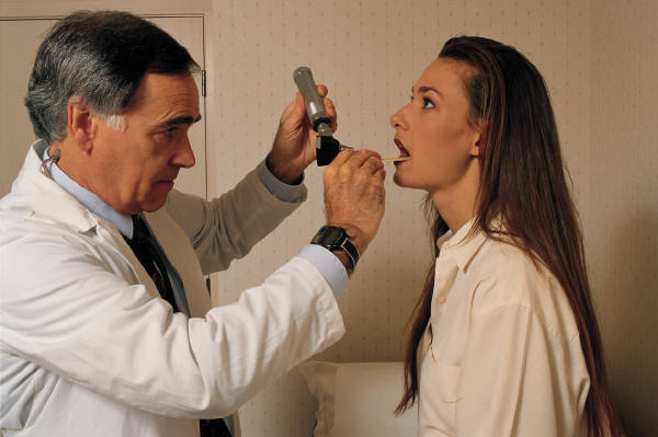
Transthoracic Echocardiogram

This is the standard, noninvasive resting echocardiogram. This procedure is
typically performed in the doctor's office or a hospital. You simply remove your
clothing and lay down on a examination table or bed. The technician will attach sticky patches or electrodes with wires to your chest and
shoulders for the EKG readings then apply a colorless gel to your chest area. He
will then rub or move a hand held echo transducer over the gelled area to makes
recordings from different parts of your chest. He does this to obtain different
views of the heart. You may be instructed to move from your back and to your
side or control your breathing while the test is being performed. The images
are viewed on the monitor while being recorded for the
physician. Sometimes a small amount of intravenous dye may be injected to
improve the image quality.
Special Preparations - None
Risks - None
Transesophageal Echocardiogram
When they can't get a good picture with the standard echocardiogram because of obstructing body mass, tissue or organs the doctor (or
technicians) have to take another path (down your throat) to get a closer look
at your heart. With a Transesophageal Echocardiogram a flexible tube with a
transducer on the end is guided down your throat and into your esophagus where
it can obtain more detailed images of your heart. In this procedure you will be
given medications to help you relax and your throat will be numbed with a spray
or gel.
Special Preparations - No eating a few hours before the
procedure. No driving after due to the
sedating medication.
Risks - Possible sore throat for a few hours after the
procedure. Possible breathing problems during the procedure (caused by the
sedation medication) but your oxygen level will be monitored during the exam.
Stress Echocardiogram

Some heart problems only show up when the heart is under stress or when you
are engaging in physical activity. For a stress echocardiogram ultrasound images
of your heart are taken immediately before and after riding a stationary bike or
walking on a treadmill. If you are physically unable to exercise medication may
be given to you to make your heart pump faster to simulate exercising.
Special Preparations - Wear comfortable clothing shoes for exercise.
Risks - Normal risks associated with exercise or medication.
Doppler Echocardiogram
Uses the Doppler Effect to help your doctor measure the speed and direction
of the blood flow in your heart. This is generally used in conjunction with both
the Transthoracic and the Transesophageal procedures.
Special Preparations - N/A
Risks - N/A
Ask A Question/Tell Your Experience
![]()


